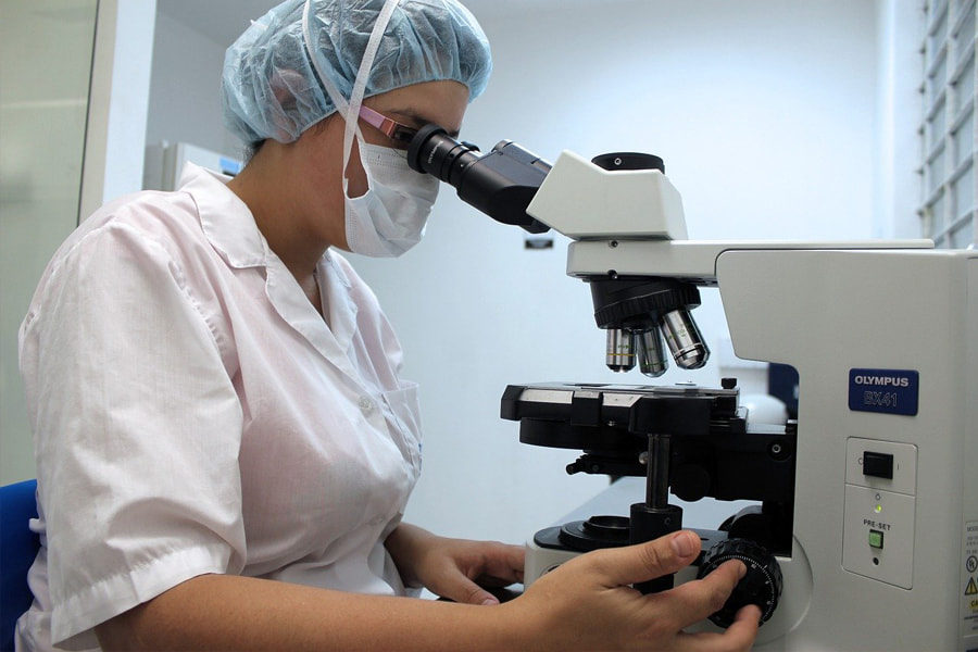
For plenty of applications in various industries, microscopes are widely in use. For various purposes, we may have to use various types of microscopes. It is through each of those types that we can fulfill our work efficiently. Obviously, different designs of microscopes are helpful in fulfilling various purposes. In this article, we are primarily focusing on the different types of microscopes that are available. Alright. Let us dive right into it. Let us discuss the different types of microscopes.
Importance of Microscopes
We utilize a lot of items in our day-to-day lives for which we have no idea about their manufacturing process. However, with a little inquiry, you may discover that microscopes are in use in various parts of the production process for such things. And microscopes are obviously critical in the production of a wide range of products. Nevertheless, we have unintentionally used microscopes on occasion. In fact, different professionals use different types of microscopes. Of course, with our content, you will be able to understand the different types of microscopes.
What are the Different Types of Microscopes?
Okay. To start with, let us list out the different types of microscopes that we can focus on. Here we go.
- Compound microscope
- Electron microscope
- Stereo microscope
- Phase-contrast microscope
- Atomic force microscope
- Confocal microscope
- Clinical microscope
- Scanning probe microscope
- Acoustic microscope
- Fluorescence microscope
- USB computer microscope
- Pocket microscope
These are the types of microscopes that we hope to focus on, throughout this article. We hope that you will be able to get a good understanding of the different types of microscopes. Keep reading!
1. Compound microscope
Under different types of microscopes, firstly, we are going to discuss the compound microscope. And now, it is time to start it off.
The term compound microscope
The term compound refers to the fact that the microscope has more than one lens. Of course, the compound microscope consists of two optical components and a system of lenses. And the optical lens and the ocular lens are the two types of lenses.
Working principle of the compound microscope
The specimen to be studied is mounted on a clear glass slide and put between the condenser and objective lenses on the specimen stage. From the base, a beam of visible light focuses onto the specimen using a condenser lens. Consequently, the objective lens detects the specimen’s light which creates a magnified picture of the specimen within the body tube termed the main image. And the eyepiece or ocular lens enlarges this image once more.
When a higher magnification is necessary, we need to turn the nose piece after low power focusing to align the higher magnification objective – usually 45X – with the lit portion of the slide. Of course, extremely high magnification is only necessary for rare circumstances. Observing a bacterial cell, for example, would need extremely high magnification. Further, an oil immersion objective lens is necessary for such situations.
Magnification of the compound microscope
To determine the overall magnification while looking at an image using a compound light microscope, we can multiply the power of the objective lens, which is often 4X, 10X, or 40X, and by the power of the ocular lens, which is typically 10X. As a result, a 10X eyepiece with a 40X objective lens will provide a 400X magnified picture, allowing the eye to observe the specimen 400X bigger than it actually is, revealing minute microscopic features.
Applications of the compound microscope
In pathology laboratories, compound microscopes are very useful as lab microscopes for identifying diseases. Also, It is in use in forensic labs to investigate and examine human cells and other materials retrieved from crime scenes in order to solve crimes. Of course, we can determine the presence or absence of metals using a compound microscope. Moreover, compound microscopes are also in use as lab microscopes in school laboratories to undertake academic investigations. Further, this is essential for studying and examining germs, bacteria, viruses, and other creatures that are ordinarily undetectable to the human eye.
Swift SW380B 40X-2500X Magnification, Research-Grade Binocular Compound Lab Microscope, Mechanical Stage, with 5.0 mp Camera and Software Windows/Mac Compatible and 100pcs Blank Slides
The binocular head on this device rotates 360 degrees for shared usage and enables rapid interpupillary distance changes without losing focus. And for added stability, you may secure the head in any position. The ocular tubes also include a 30-degree ergonomic tilt to relieve neck and eye strain. The microscope also comes with two sets of interchangeable 10X and 25X glass ocular eyepieces. This is a fantastic microscope to consider if you are seeking one.
2. Electron microscope
Now, under the different types of microscopes, we are going to talk about the electron microscope. But initially, let us understand what an electron microscope is.
What is an electron microscope?
As a source of light, an electron microscope employs a beam of accelerated electrons. And it is a kind of microscope with great picture resolution and the ability to magnify things down to nanometers. Further, these have used a phosphorescent screen and controlled the utilization of electrons in a vacuum. Ernst Ruska (1906 to 1988), a German engineer and academic professor, created the first electron microscope in 1931. Also, the basis for the modern electron microscopes is also the same prototype that Ernst Ruska created.
Working principle of the electron microscope
To gain information about structure, morphology, and composition, electron microscopes employ signals generated by the interaction of an electron beam with the material. The electron cannon produces electrons. The electron beam focuses on the specimen and converts it into a narrow tight beam by two pairs of condenser lenses. An accelerating voltage – typically between 100kv and 1000kv – is necessary to be available between the tungsten filament and the anode to transport electrons down the column. The specimen to be inspected has been thinned to a razor’s edge. Thinner than those used in optical microscopes by at least 200 times. And ultra-thin sections with a thickness of 20-100 nm are cut and put on the specimen holder.
The electrons are dispersed as the electronic beam goes through the specimen, depending on the thickness or reflective index of various regions of the specimen. Because fewer electrons reach that area of the screen, the denser portions of the specimen scatter more electrons and look darker in the picture. Transparent portions, on the other hand, are brighter. The electron beam leaves the specimen and travels to the objective lens, which has a high magnification power and creates the intermediate magnified picture. After that, the ocular lenses enlarge the final picture.
Parts of the electron microscope
The electron microscope is a tall vacuum column that’s standing vertically. And there are a few predominant parts of an electron microscope that we can consider in this regard. Firstly, we can consider the electron gun. It is the electron gun that generates electrons through a heated filament.
And then we have the electron magnetic lenses. Here, the electron beam focuses on the specimen by the condenser lens. And these electrons are formed into a thin, tight beam by a second condenser lens. The electron beam exiting the specimen goes via the objective lens, the second of the magnetic coils. This has a high magnification, resulting in an intermediate magnified image. The final enlarged picture is created by the third pair of magnetic lenses, known as projector (ocular) lenses. Each of these lenses works as an image magnifier, preserving a high degree of detail and resolution throughout the process.
Also, we have the specimen holder as an important part of the electron microscope. It is an extremely thin film of carbon or collodion held by a metal grid. Finally, we have the image viewing and recording system. This projects the final image to a fluorescent screen. Below the fluorescent screen is a camera for recording the image.
Applications of the electron microscope
Okay. Now, let us take a look at the applications of the electron microscope. Mainly, electron microscopes are in use for the studies of the structure of a wide range of biological and inorganic specimens. Microorganisms, cells, huge compounds, biopsy samples, metals, and crystals are among them. Electron microscopes are in use in industrial research for quality control and failure analysis too.
Modern electron microscopes capture images using specialized digital cameras and frame grabbers, resulting in electron micrographs. Microbiology owes much of its advancement to the creation and improvement of the electron microscope. The study of microorganisms such as bacteria, viruses, and other pathogens has greatly improved illness treatment.
3. Stereo microscope
The next type of microscope that we are going to consider is the stereo microscope. Let us go through it now. Sometimes, we refer to the stereo microscopes as dissecting microscopes as well.
It is a low magnification digital optical microscope with a magnification range of 5X to 250X. The magnification is obtained by light reflected off the specimen’s surface rather than light reflected on the specimen itself. The dissecting microscope’s principal function is to observe and qualitatively assess dissected specimen material. In 1977, Cherudin D’orleans created the first stereo microscope, which consisted of a tiny microscope with two distinct eyepieces and objective lenses.
Working principle of the stereo microscope
The two types of light channels employed by microscope objectives and eyepieces determine the operating principle of the stereo (dissecting) microscope. Each light path delivers a unique perspective. They have a top light that is for dissecting and a bottom light that is for viewing the photographs. The design of two eyepieces (binocular stereoscope) allows for this illumination, each presenting a separate sort of light pathway and offering an easy viewing region.
It displays the photos live on a computer monitor screen in 3-dimensional visuals because it is a digital microscope. They also allow for a very close examination of minute specimens such as insects, which appear as much bigger pictures than the sample’s true size. Macro photography is the term for this type of magnification. The surface is evaluated in 3D and the image is stored in complicated samples.
Two magnification mechanisms are available in the stereo microscope. Fixed (primary) magnification is a type of magnification in which two objective lenses are used to create a degree of magnification. And the zoom (pancreatic) magnification delivers continuous magnification across a wide range of distances by employing auxiliary objectives that boost overall magnification based on various circumstances. Changing the eyepiece lenses allows you to choose between zoom and fixed magnifications.
Types of stereo microscopes
Stereo microscopes also have some types that we can consider here. What are they? Let us take a look.
- Stereo zoom dissecting microscope
- Digital tablet dissecting microscope
- Stereo zoom boom stand microscope
- Dual power dissecting microscope
- Single power stereo dissecting microscope
- Single magnification handheld pocket microscope
You would like to know about these types for sure. Do not worry. We are willing to tell you about them.
Stereo zoom dissecting microscope
Stereo zoom dissecting microscopes feature a zooming range of 6.7x to 45x. And they are triangular or binocular dissecting microscopes. You can connect it to a digital camera, which will capture pictures of the visuals you’re looking at. These microscopes have a 360-degree rotation and dual-LED illumination. By adding supplemental objectives or other eyepieces, you may change the magnification too.
Digital tablet dissecting microscope
These are some of the high-tech dissecting microscopes available. They have a touch panel LCD tablet camera with a magnification range of 6.7x to 45x and a continuous magnification of 6.7x to 45x. And they have supplemental eyepieces that you can adjust in magnification range. To change the magnification, you can add additional lenses to the objectives too. They have a 5.0-megapixel camera that can capture photographs and movies and save them immediately to the tablet or through a USB connection. At the top and bottom of the microscope, there are built-in LED illuminators that function independently.
Stereo zoom boom stand microscope
You can use these for inspecting huge samples. These have a large base and the largest stage. They have a zooming range of 6x to 45x, which you can adjust upward by adding additional lenses or eyepieces.
Dual power dissecting microscope
Typically, this type includes a 10x and 30x dual-powered dissecting microscope with a 360* rotation capability for focusing and viewing. And you can change the visual magnification by rotating the lenses. It also has a high-intensity LED light ring that illuminates the whole surface. When studying bigger specimens, this microscope’s flexible stand allows you to raise to a height of up to 6 feet.
Single power stereo dissecting microscope
They offer modest magnification powers ranging from 10x to 40x, as well as 45-degree tilted eyepieces. These microscopes also include diopter settings ranging from 50mm to 70mm.
Single magnification handheld pocket microscope
This is a light-free, single-powered portable microscope with two magnification capabilities. This is very easy to use for examining specimens. Additionally, it is portable due to its small size.
AmScope SE305-P Binocular Stereo Microscope, WF10x Eyepieces, 10X and 30X Magnification, 1X and 3X Objectives, Upper and Lower Halogen Lighting, Reversible Black/White Stage Plate, Pillar Stand, 120V
This stereo microscope comes with two 10x18mm widefield eyepieces, 1x and 3x objectives, upper and lower halogen lighting, two stage plates, and pillar support. To make seeing simpler for youngsters, the binocular viewing head has a 55-75mm interpupillary range and a 45-degree tilt. The WF10x18mm eyepieces combine with the 1x and 3x objectives to give 10x and 30x magnification and a larger working distance for studying large-scale specimens that require handling or restoration. To generate high-resolution photographs and sharp images, the optical glass lenses have full coatings.
4. Phase-contrast microscope
Frits Zernike, a Dutch scientist, was the first to invent the phase-contrast microscope idea. The phase-contrast microscope is an optical microscope method that uses brightness to transform phase shifts in light traveling through a transparent object into picture alterations. Unstained live cells absorb nearly no light, therefore phase contrast microscopy is in use to examine them. In other optical microscopes, poor light absorption causes extremely minor changes in the intensity distribution in the picture, making the cells scarcely visible or not visible at all.
Working principle of the phase-contrast microscope
The tungsten-halogen lamp produces moderately clear light, which goes through a collector lens and focuses on a specific annulus in the sub-stage condenser front focal plane. Wavelengths flowing through the annulus illuminate the specimen and either pass through undeviating or are diffracted and delayed in phase by features and phase gradients in the material. The phase plate separates the un-deviated and diffracted light gathered by the objective at the rear focal plane and focuses it at the intermediate image plane to generate the final phase-contrast picture seen via the eyepiece.
AmScope PCS Phase Contrast Kit for Compound Microscopes
For compound microscopes, this is a complete phase contrast kit. Three-phase contrast plan objectives (10x, 40x, and 100x), three-phase contrast condensers, and a CT lens are in this. The condenser has a 37mm mounting size (in diameter). The Plan goals have a 20mm thread (in diameter).
5. Atomic force microscope
Under the different types of microscopes, we are now going to consider atomic force microscopes. Let us go ahead with it.
What is an atomic force microscope?
The atomic force microscope (AFM) is a scanning probe microscope that is in use to measure qualities such as magnetism, height, and friction. You can measure the resolution in nanometers, far more precise and effective than the optical diffraction limit. For measuring and collecting data, the AFM uses a probe that includes contacting the surface with the probe. When the scanning probe microscope raster scans the probe over a piece of the material while simultaneously measuring its local characteristics, it creates a picture.
The AFM also includes piezoelectric components, which are electric charges that build in solid objects such as DNA, biological proteins, crystals, and other materials, allowing for microscopic, precise movement during scanning in response to an electric command. Following the creation of the scanning tunnel microscope for IBM Research in Zurich in1982, the IBM scientists developed the AFM In 1986, the Atomic Force Microscope was in use for the first time in an experiment, and in 1989, it was available for commercial usage.
Parts of an atomic force microscope
The AFM can measure force interactions using a variety of approaches, including thermal, electrical, and magnetic force. The AFM contains various elements that govern its activities in order to make these measurements possible.
Mainly, these have modified tips to detect the sample surface and deflect. Adjustments to the software that is used to image the samples. And these have a feedback loop control system. A laser deflector is used to manage the force interactions and tip locations in a feedback loop. The laser bounces off the rear of the cantilever and the tip, and when the tip interacts with the sample’s surface, the laser’s location on the photodetector is employed in the feedback loop to monitor the sample’s surface and quantify it.
A laser beam deflection mechanism is used to construct the AFM. The laser is reflected in the sensitive detector from the rear of the Atomic Force Microscope lever. These are constructed of silicon compounds and have a tip radius of around 10 meters. The force interactions that contribute to the picture created are crucial to the AFM’s operation. When the stiffness of the cantilever is known, the forces are calculated using the deflection lever.
Applications of the atomic force microscope
AFM microscopy is in use in a range of fields, including natural science and other fields. Mainly, this is in use in the detection of atoms in samples. And for analyzing atom-to-atom force interactions also, this is in use. Also, this is useful for the study of atoms’ changing physical characteristics. Further, the structural and mechanical features of protein complexes and assemblies, such as microtubules, can also be easily investigated. Importantly, these are in use to distinguish cancerous from non-cancerous cells. Observing and distinguishing nearby cells, as well as their form and cell wall stiffness is also possible with this.
6. Confocal microscope
Under the types of microscopes, it is now about the confocal microscope. Let us take a look at this type in detail now.
History and origin
Marvin Minsky, a Harvard University neuroscientist, invented the confocal microscope in the 1950s with the goal of visualizing the neural network without straining the tissues. However, owing to a lack of a suitable light source and a computerized method to store the enormous data, the confocal microscope did not go farther at the time. This further expanded the confocal microscopy idea in the late 1960s, with the invention of the multiple beam confocal microscope. This innovative invention utilized the Nipkow spinning disk, which was used to analyze unstained brain tissues and ganglion cells.
The concept of confocal microscopes has evolved throughout time as science, technology, computers, and digital image management and processing have progressed. In 1987, the first commercial confocal microscope was created, featuring improved optics and electronics, as well as strong lasers and great scanning efficiency. The current confocal microscope incorporates every technological and mechanical component imaginable, including optical components that execute the principal purpose of the design through the use of electrical detectors, a computer, and a laser system.
The confocal microscope’s operation is the result of all of its components working together to form an electronic image. These microscopes have previously been used to study molecules, microbial cells, and tissues.
Working principle of the confocal microscope
Traditional (wide-field) microscopes illuminate a vast region of a specimen by using multiple wavelengths from a light source. This approach creates blurry, unclear, and congested pictures because cell sample images are taken from all angles without a focus point. To circumvent these problems, the confocal microscope idea is ideal. The entire specimen gets light and emits it in a wide-field or fluorescent microscope, which is detected by a photodetector on the microscope. The principle and operation of a confocal microscope, on the other hand, is based on lighting a focus point on the specimen.
Prior to the examination, a specimen is stained with fluorochrome. When a beam of light is focused on a specific location on the floro-chromatic specimen, the objective lens focuses the illumination to a plane above the objectives. On the focal plane above the objective, there is an aperture. The primary purpose of this opening is to prevent stray light from reaching the specimen. The illumination point is 0.25 to 0.8um in diameter and 0.5 to 1.5 um depth with brilliant intensity, as specified by the objective numerical aperture.
The specimen is frequently found between the camera lens and the plane of focus, which is the perfect point of focus. The microscope’s laser scans a plane on the specimen (either via beam scanning or by rotating the stage) (stage scanning). The illumination will subsequently be measured by a detector, resulting in a picture of the optical section. Several optical portions are scanned and stored as data in a computerized system, resulting in a three-dimensional picture. The image may be quantified and measured.
Parts of the confocal microscope
In this sector, we discuss the important parts of the confocal microscope. Firstly, we have the objective lens. Then, we have the out-of-focus plane and the in-focus plane. We also have the beam splitters, detector, confocal pinhole or aperture, laser, and oscillator mirrors too.
Types of confocal microscopes
There are a few types of confocal microscopes that we can discuss in this regard. Let us first list them out.
- Confocal laser scanning microscope
- Spinning disk
- Dual spinning disk
- Programmable array microscope (PAM)
You would surely be curious to know about each of these. Let us go through them descriptively.
Confocal laser scanning microscope
In this microscope, what happens is, by scanning and de-scanning, numerous mirrors scan along the X and Y-axis of the object. The picture is then sent into the detector through a pinhole.
Spinning disk
This is a confocal microscope that employs numerous moveable apertures (pinholes) on a disc to scan for spots of light in a parallel fashion across a given plane over a lengthy period of time. We refer to this as the Nipkow disk as well. When compared to a laser scanning microscope, the longer the exposure period, the less excitation energy is required for illumination. Phototoxicity and photobleaching are reduced when the excitation energy is reduced, hence it is mostly used for imaging cells.
Dual spinning disk
This functions similarly to a spinning disk confocal microscope. The difference is that it has a second disk with pinholes on it. The microlens collects a broad spectrum of light and concentrate it into each pinhole, minimizing the quantity of light obstructed by the spinning disk. The confocal microscope with improved microlens is substantially more sensitive than the spinning disks as a result of this change.
Programmable array microscope (PAM)
In this microscope, a spatial light modulator is used. It is an object that imposes a form of spatially variable modulation on a beam of light, abbreviated as SLM. A collection of moveable apertures (pinoles) with arrays of pixels of opacity, reflectivity, or optical rotation are used in the SLM. A charge-coupled device (CCD) camera captures the image captured by the SLM’s microelectrochemical mirrors.
Applications of the confocal microscope
Biochemical sciences, cell biology, spectroscopy, genetics biology, nanoscience, developmental biology, and quantum optics are just a few of the domains where confocal microscopes are in use.
For measuring and qualitatively examining the endothelial cells of the cornea, the confocal microscope is in use to study ocular corneal infections in biomedical sciences. During keratomycosis infection, it is also in use to detect the presence of fungal elements in the corneal stroma. Additionally, it is in use for fast diagnostic and treatment response. And in the pharmaceutical industry, to maintain thin-film medicines, allowing quality and consistency of medication delivery, these microscopes are in use.
Advantages of the confocal microscope
As a confocal microscope studies the image from one optical point to another, it improves the image’s outcome. As a result, light from other regions of the specimen is not dispersed. And the particular focal point is displayed and recorded too. Of course, you can use the confocal microscope to examine both living and dead cells. Further, It is possible to gather serial optical portions using it. In fact, the confocal microscope illuminates the exact points of interest consistently. The zoom factor is used by confocal microscopes to modify their magnification electronically without changing the objectives. And the confocal microscope produces a series of three-dimensional pictures.
Disadvantages of the confocal microscope
Nevertheless, there are certain disadvantages of confocal microscopes too. One is that the excitation wavelengths of the confocal microscope are restricted, with very thin bands. Also, the ultra-violet light emitted by confocal microscopes is costly to manufacture. Moreover, confocal microscopes are expensive to manufacture and acquire. Although there are these drawbacks, there are plenty of advantages of confocal microscopes.
Basic Confocal Microscopy 2nd Edition, Kindle Edition
This is a book by W. Gray (Jay) Jerome and Robert L. Price. Basic Confocal Microscopy, Second Edition expands on the previous edition’s success by maintaining the same format while also addressing pertinent modifications and new discoveries in this still-emerging area. This format is based on several of the authors’ Confocal Microscopy Workshop, which has been given by many of them for almost 20 years and is still a popular workshop for learning the fundamentals of confocal microscopy.
7. Clinical microscope
Clinical microscopes are required for biological research conducted by clinicians, lab supervisors, and medical workers. For clinical university and institutional applications, clinical microscopes, also known as medical microscopes, are necessary. Clinical or medical microscopes come in a variety of shapes and sizes and are available from a variety of manufacturers.
Types of clinical microscopes
Alright. Now, let us get to know the types of clinical microscopes that are available. Here they are.
- Biotech tissue culture microscope
- Epi-fluorescence microscope
- Gout microscope
- IVF / ART microscope
You would like to know about them in detail. Let us tell you. Keep reading!
Biotech tissue culture microscope
This is one type of clinical microscope that we can talk about. These are mainly for tissue culture work in demanding atmospheres.
Epi-fluorescence microscope
This is one of the most popular optical microscopes on the market. This equipment may be used in conjunction with high-quality Japanese optics and a range of filter cubes to achieve various degrees of resolution.
Gout microscope
Medical physicians utilize gout microscopes, which are designed specifically for recognizing gout or CPPD (false gout) crystals suspended in synovial fluid. These are very useful for this field.
IVF / ART microscope
These microscopes are made to do exact reproduction work. Depending on whatever component of the reproductive system is the basis of the study, different microscopes are in use.
8. Scanning probe microscope
Scanning probe microscopes enable high image magnification for observation of three-dimensional formed objects, providing researchers with imaging capabilities for the future. When you stimulate or handle the specimens, this results in much better photos as well as specimen qualities, responsiveness, and reaction or non-action.
History and origin
In 1986, Rohrer and Binning received the Nobel Prize in Physics for their achievement in transferring scanning probe microscopy technology from the drawing board to the laboratory. Scanning probe technology at the microscopic level is now a standard analysis tool for research and development in both academic and industrial facilities, including physics, biology, and chemistry.
Working principle of the scanning probe microscope
A pointer, electrically charged probe is there to trace the surface of a specimen, similar to how an old record player made music by a needle tracing the grooves of an LP. The SPM probe, unlike a record player needle, does not touch the surface but instead tracks the specimen nanometers above it.
The probe may also be in use to interact with a specimen, allowing researchers to see how a material attracts or repels, as well as how it reacts to electrical currents. Non-conductive specimens can be manipulated and viewed using SPM technology since they can work in a broad range of settings.
Advantages of scanning probe microscopes
Scanning Probe Microscopy allows researchers to observe specimens in a wider range of conditions while using the same microscope and specimen, cutting down on the time it takes to prepare and examine specimens. With minimal effort and modification, specialized probes, upgrades, and adjustments to scanning probe devices continue to deliver quicker, more efficient, more revealing specimen pictures.
Disadvantages of scanning probe microscopes
One of the disadvantages of scanning probe microscopes is that this creates pictures in black and white or grayscale. That might exaggerate a specimen’s real form or size in some cases. Computers are in use to adjust for exaggerations and provide real-time color pictures that offer researchers real-time data on cellular interactions, harmonic responses, and magnetic energy.
9. Acoustic microscope
Acoustic microscopy is a method that exhibits science and technology’s developments and achievements. It helps scientists to better evaluate and monitor micro-environments, as well as expand research, generate the potential for discovery, and allow for a deeper examination of a specimen. Scientists can collect more precise data, verify or refute a hypothesis, develop a better grasp of the invisible world around us, and explore novel cures, therapies, or products to make everyday life safer and healthier by improving their capacity to analyze a sample.
Advantages of the acoustic microscope
A standard microscope can only view the top of the material, maybe even the subsurface, however, an acoustic microscope can zoom in on a single location and receive pictures from deeper layers. Another fascinating feature of the acoustic microscope is its ability to identify carbon fiber from other materials that a producer may employ to strengthen a product. Also, the use of ultrasonic waves allows for the determination of a focus point by reducing diffraction, allowing for more accurate findings and data while maintaining the integrity of the sample.
Disadvantages of the acoustic microscope
Acoustic microscopy, as interesting as it is, has several drawbacks, mostly due to the naturally occurring structure of sound waves. Slow processing time, too high a sensitivity to delaminations, expense, and failure to adjust fast to economic changes are all issues that technology has failed to solve.
10. Fluorescence microscope
By altering the formation of an image, you can use these microscopes to observe different elements of the material. Fluorescence microscopes interact with dyes by using certain hues of light. Certain structures may be separated and seen with their particular colors when the dyes become lit. This microscope is useful to look at individual proteins within a cell. A camera is common with this microscope to collect pictures.
AmScope 40X-1000X Upright Fluorescence Microscope with Rotating Multi-Filter Turret + Ultra-sensitive CCD Camera
So, this upright fluorescence microscope has a six-filter turret with transmitted and reflected light. The microscope has a Koehler diascopic and episcopic illumination system, as well as an infinity-corrected optical system with a magnification range of 40X to 1000X. Moreover, in the powerful 100W wide-spectrum mercury-vapor episcopic illuminator, a rotating filter-selector contains up to six fluorescence filter-blocks; already included are ultraviolet, violet, blue, and green excitation blocks. Furthermore, the substage illuminator uses a 30W halogen lamp with field-lens and diaphragm for even, high-contrast light.
11. USB computer microscope
A computer microscope or a computer-connected microscope is another name for a USB computer microscope. And it is a microscope that plugs into a USB port on a computer or television and uses CMOS sensors. Of course, instead of staring at the specimen through an eyepiece, the viewer views it on a computer display or a television screen. And it is just a webcam with a macro lens with it.
Advantages of a USB computer microscope
The majority of these devices are portable, allowing viewers to examine specimens that would not fit beneath the lens of a regular microscope. Also, there is the ability to save the photographs as picture files with ease. And the user may even save the photos as a video clip on some devices. Further, in other apps, you may analyze or alter the photographs you’ve saved. Of course, this allows the spectator to zoom in on details that may otherwise go unnoticed, as well as edit the images for creative or entertaining purposes. These are the major advantages that you can get through this type of microscope.
Disadvantages of a USB computer microscope
All the same, there are some drawbacks of this model too. One is the low-level magnification. And the other is the issue with insufficient illumination on certain occasions.
7 inch LCD Digital USB Microscope with 32G TF Card, Micsci 1200X Magnification 12MP 1080P Handheld Camera Video Recorder, PC View, Rechargeable Battery, Fill Lights for Coins PCB Soldering Circuit Board
So, in this set, 8 brilliant adjustable LED lights and 2 additional auxiliary lights are available. When the light source is insufficient, it may turn on the light, providing outstanding detail and clarity, and allowing you to take images or video in the woods and other dark areas. Also, you can easily move up and down the bracket, making it very easy to change the object’s distance. The stand’s angle is also adjustable, allowing users to select the most comfortable and appropriate viewing angle from the display.
12. Pocket microscope
Children, students, and scientists can examine items in amazing detail both outdoors and inside with a portable microscope. Moreover, some of these microscopes are as little as an ink pen and produce detailed up-close views of objects and bigger single-celled animals. Due to this fact, they are compact, sturdy, and portable. And because of that, many of these hand-held microscopes do not require batteries and may work with natural light to provide high-definition images with no blurring. You can hold in the hand, move over a big object, or carry in a pocket, portable microscopes, unlike traditional microscopes that require a firm platform to sit on. Further, portable and pen-sized microscopes with magnification ranges of 25x to 100x utilize batteries and contain an LED light. Some of these microscopes also include a focus adjustment.
Applications of the pocket microscope
You can use many of these pocket microscopes in a variety of settings, including the classroom, and some come with rubberized eyepieces for added comfort and safety, particularly when used by young children. Moreover, the majority of these microscopes have no moving components and are ideal for introducing youngsters to the hidden world around them.
Furthermore, emergency medical technicians, trauma and emergency room practitioners, as well as scientists and enthusiasts, use the device. Of course, pocket microscopes are extremely useful in the manufacturing industry for detecting flaws in electrical components, metals, optics, glassware, and structural flaws in equipment.
Carson Pocket Micro 20x-60x LED Lighted Zoom Field Microscope with Aspheric Lens System (MM-450), Blue
So, if you are looking for a pocket microscope among the different types of microscopes, we think that this would be a good one for you to go for. Further, for factors like ease of use, value for money, and sturdiness, this has received good ratings on Amazon. Also, this has received favorable reviews from its customers. So, we think that this would surely be a good one for you to go for.
Conclusion
So, in this article, we primarily focused on the different types of microscopes. Consequently, we hope that by now, you have a good understanding of the different types of microscopes that are available. Furthermore, if you wish, you can read about the uses of microscopes too.