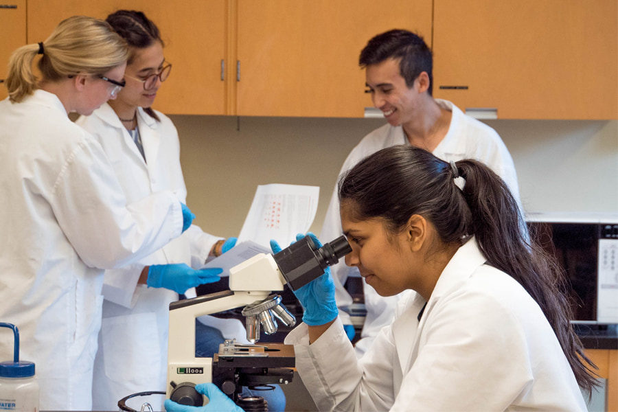

Microscopes are in use in a wide range of industries and for a vast range of applications. The invention of microscopes took place long ago. Now, for diverse purposes, we may need to use a variety of microscopes. Each of those areas can help us do our work quickly. Microscopes exist in a wide range of forms and sizes, and they are helpful for a wide range of jobs. Microscopes are quite useful in all of these fields. One special purpose of using microscopes is to view bacteria. In this article, we are going to talk about bacteria under microscopes. We are pretty sure that through this article, you will be able to get a good understanding of bacteria under microscopes.
About Microscopes
In our daily lives, we use many goods for which we have no knowledge of the production process. However, little research may reveal that microscopes are in use in many stages of the manufacturing process of a lot of items.
Microscopes are incredibly valuable in a wide range of study fields as well as in everyday life. Of course, microscopes are in use in a wide range of industries and laboratories, including biology, chemistry, microbiology, fiber research, and drug development, to name a few. Microscopes, of course, are equipment for seeing and examining objects and specimens that are too small that we cannot see with the human eye. Despite the fact that microscopes have been around for millennia, technology has progressed substantially. Today, there are many different types of microscopes that are in use in a range of vocations and research, as well as in homes and schools.
An optical microscope with a single lens is the most basic of all. Simple microscopes include magnifying glasses and jeweler’s loupes. A compound microscope is an optical microscope with two lenses. The two basic elements of a compound microscope are the objective, which holds the lens near the specimen, and the eyepiece, which holds the lens near the spectator. A contemporary compound microscope additionally has a light source (either a mirror to gather exterior light or a light bulb to supply interior light), a focusing mechanism, and a stage (a surface on which you can hold the object that you need to examine.)
OMAX 40X-2000X LED Binocular Compound Lab Microscope w/ Double Layer Mechanical Stage + Blank Slides, Cover Slips, & Lens Cleaning Paper, M82ES-SC100-LP100
This compound biological microscope has magnification levels of 40X to 2000X. The microscope’s objectives are 5X, 10X, 40X, and 100X. On both sides are coaxial coarse and fine focus knobs. This ergonomic design allows you to effortlessly change the focus to get the results you want. Transmitted illumination, such as an LED light that may be changed in intensity based on the scenario, is also included. It also has a far longer lifespan than a standard halogen bulb.
What is Bacteria?
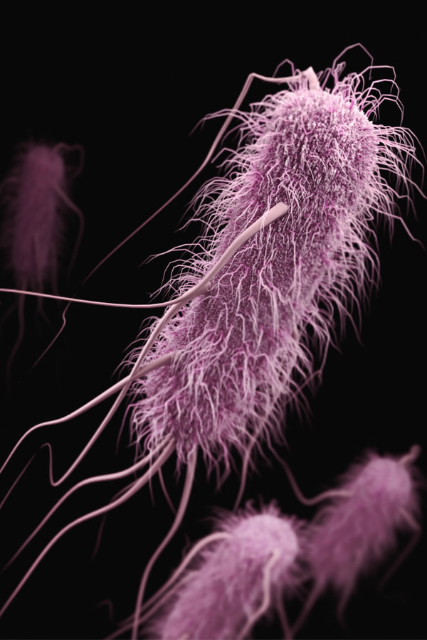
Bacteria are microscopic living creatures that we can find in large quantities in practically any place on the planet. From the depths of the earth to the intestines of humans. We can only see bacteria under the magnification of a microscope since they are too tiny to see with the naked eye.
Most bacteria have a diameter of 0.2 um and a length of 2-8 um, and come in a variety of forms, from spheres to rods and spirals. Bacteria only seem colored when they are part of a colony; lone bacteria appear clear. The bacterial cells will drift in and out of focus at high magnification, especially if the water layer between the cover glass and the slide is too thick. It needs a knowledgeable individual to tell the difference between germs and little dust and grit on a slide. Because certain bacteria are existing in clusters, it is difficult to see individual cells.
We can divide bacteria into five types considering their fundamental shapes: spherical (cocci), rod (bacilli), spiral (spirilla), comma (vibrios), and corkscrew (corkscrew bacteria) (spirochaetes). They might be single cells, pairs of cells, chains, or clusters.
Bacteria exist in many types of environments on Earth, including soil, rock, seas, and even polar snow. Some creatures, including plants and animals, including humans, live in or on other organisms. In the human body, there are around ten times as many bacterial cells as there are human cells. We can discover many of these bacterial cells in the lining of the digestive tract. Bacteria are there in the soil or on decaying plant debris, where they play a vital role in nutrient cycling.
National Optical 40X-1000X Compound Microscope Set with Slides for Students and Kids Biology Cordless Beginner Microscope All Metal
Thanks to the 4X, 10X, and 40X objectives, the magnification of this optical microscope span from 4X to 1000X. Furthermore, the visual clarity and sharpness are outstanding. Furthermore, chromatic aberrations and edge blur are compensated for with this microscope. The head of this monocular is tilted 45 degrees and swivels 360 degrees. As a consequence, the eyes and neck are less stressed. Regardless, it has a highly precise coaxial focusing mechanism. It’s quite simple to use as a result of all of this.
Preparing bacteria samples for viewing
Collect and drip a tiny amount of distilled water over the slide with a clean dropper or inoculating loop. Then, with the distilled water, drip a little quantity of bacterial culture. To mix the bacteria with the distilled water, sweep the inoculating loop over the glass slide. Place the slide on a drying rack to dry before viewing, or cover the slide with a coverslip to watch the bacteria in action.
Samples may need to be cultivated and Gram-stained in advance due to the tiny size and sometimes translucent nature of bacteria. The concentration of cells in a sample increases as bacteria are cultured. To Gram-stain the culture, soak it for one minute in crystal violet, methylene blue, or safranin, then gently remove the excess stain using water or an absorbent cloth.
80-2000X Optical Microscope, Metal Body, 2 WF Oculars, Dual-illuminators System, US Plug, Full Accessories for Kids Students Beginners
When using this microscope, choose a low magnification combination (80X) to study the entire specimen clearly, and a higher magnification combination to analyze the specimen in detail (max 2000X). Images of the same sample captured at different magnifications may also be compared. Use wide-field eyepieces made of high-quality all-optical instruments to view brighter and clearer. With achromatic goals, you may view the most realistic microbiological environment.
Bacteria under the microscope
Observing bacteria under microscopes is similar to observing anything else under a microscope. Prepare a bacterium sample on a slide and place it on the stage of the microscope. Change the objective lens after adjusting the focus until the bacteria appear in the field of view. Before going on to the next objective lens, repeat the focus adjustments and continue until the appropriate magnification is achieved.
All the same, we are ready to give you some tips on viewing bacteria under microscopes. Just go ahead reading! There are some interesting facts coming up.
Preparing a sample
Obviously, you can begin by using a clean slide and distilled water to produce a bacterium sample. Make sure your dropper is totally clean before putting the water on the slide. A soiled dropper may easily distort your findings and make finding what you are searching for much more difficult. After a few drops of distilled water have been poured on the slide, add your bacteria culture. Because bacteria are just a few millimeters long, Gram staining may be beneficial in some cases. Methylene blue or safranin can be used to stain your cell culture for Gram staining. Your bacteria will be easier to observe under the microscope as a result of this.
Check with the lens sizes
A lens with at least a 400x magnification will be necessary to view bacteria swimming. Bacteria can be seen in breathtaking detail at 1000x magnification. However, as the magnification increases, keeping them in focus while they move becomes more challenging. Start small and focus before moving up to the next degree of magnification, regardless of the greatest magnification you intend to utilize. Skipping steps will make it much more difficult to concentrate on larger numbers.
Safety practices
Prepared slides are the way to go for classroom experiments or fun at home, especially with younger pupils. In the aim of discovering an intriguing sample, avoid utilizing damaged food or bodily fluids. When working with bacteria, always use gloves. In fact, wearing gloves whenever you use a microscope is a smart idea to avoid dirt and body oils from clouding your vision.
Take photos
You might want to take your experiment to the next level and photograph your topic if you have mastered the art of detecting and recognizing different varieties of bacteria. Images from a digital microscope may be projected and saved straight to a computer using microscope cameras. Photographing microorganisms may generate spectacular, artistic effects, which may pique your interest in a whole new pastime. There are a number of microscope cameras and accessories available to assist you in capturing a wide range of photographs. You may alter the photographs into vibrant and powerful compositions using an equally huge number of image editing tools.
AmScope GM400TZ-10M Digital Trinocular Gemology Stereo Zoom Microscope, WH10x Eyepieces, 3.5X-90X Magnification, 0.7X-4.5X Zoom Objective, Halogen, and Fluorescent Lighting, Inclined Pillar Stand, 110V-120V, Includes 0.5X and 2.0X Barlow Lenses, 10MP Camera with Reduction Lens, and Software
A lengthy working distance allows users to work with or touch huge goods like jewels and jewelry, while a 10MP camera with a reduction lens and USB 2.0 output let them snap and display photos on a computer or projector. There’s also a trinocular viewing head with 10x widefield high-eyepoint eyepieces, adjustable interpupillary distance, fixed 45-degree vertical inclination to relieve eye and neck strain, and 360-degree rotation for sharing.
Final Thoughts
This article mainly focused on viewing bacteria under microscopes. Of course, there are so many other uses of microscopes too. We think that you have a general idea of how you should inspect bacteria under microscopes, by now. So, now, you can go ahead and view bacteria under microscopes. The microscopes are all yours to do so.